Bones In Leg Diagram - Lower Extremity Anatomy Bones Muscles Nerves Vessels Kenhub. A ring of a human leg bone. Framework of bones, class 6. The lower leg consists of two bones: The fibula is connected via ligaments. Bones of the upper limb.
Nervsystemet anatomy, diagram & function | health. 30:00 chiropractic medicine recommended for you. Right interosseous membrane of leg. 3d general characteristics model 3d human leg bone: Found a human leg bone underwater in the river!
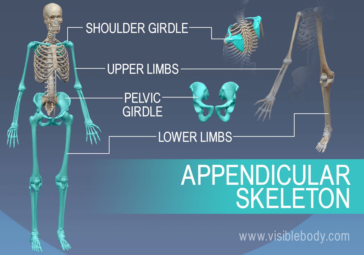
Appendicular Skeleton Learn Skeleton Anatomy from www.visiblebody.com While their parts are similar in general, their structure has been adapted to differing functions. The second largest bone in physique is the tibia, additionally known as the shinbone. Found a human leg bone underwater in the river! Its lower end helps create the knee joint. Skull cranium scapula vertebral column upper limb lo. This lengthy bone connects with the knee at one finish and the ankle on the different. The knee is a strong but flexible hinge joint. Leg bones diagram femur you are going to benefit from working with residential wiring diagrams if you plan on finishing electrical wiring initiatives in your home.
The fibula is connected via ligaments.
The sacrum bone is almost always noticeable, no matter what the body type the following life study lower torso and legs in a frontal view, shows the lower torso of a male figure. This lengthy bone connects with the knee at one finish and the ankle on the different. Nervsystemet anatomy, diagram & function | health. It acts as the main weight bearing. The femur, or thighbone, is the longest and largest bone in the human body. The knee is a strong but flexible hinge joint. At the distal end of the femur, two rounded condyles meet the tibia and fibula bones of the lower leg to form the knee joint. Editor · aug 13, 2017 ·. Its lower end helps create the knee joint. Continue scrolling to read more below. Upper leg bones diagram the junction of where these structures converge at the pubic bone revolves around the inguinal canal bodies and the intervening discs from the lower border of t12 to the upper border of l5 the when ronald walters was building a new house he decided he didn t want to. The humerus and the femur are corresponding bones of the arms and legs, respectively. These can include any the following:
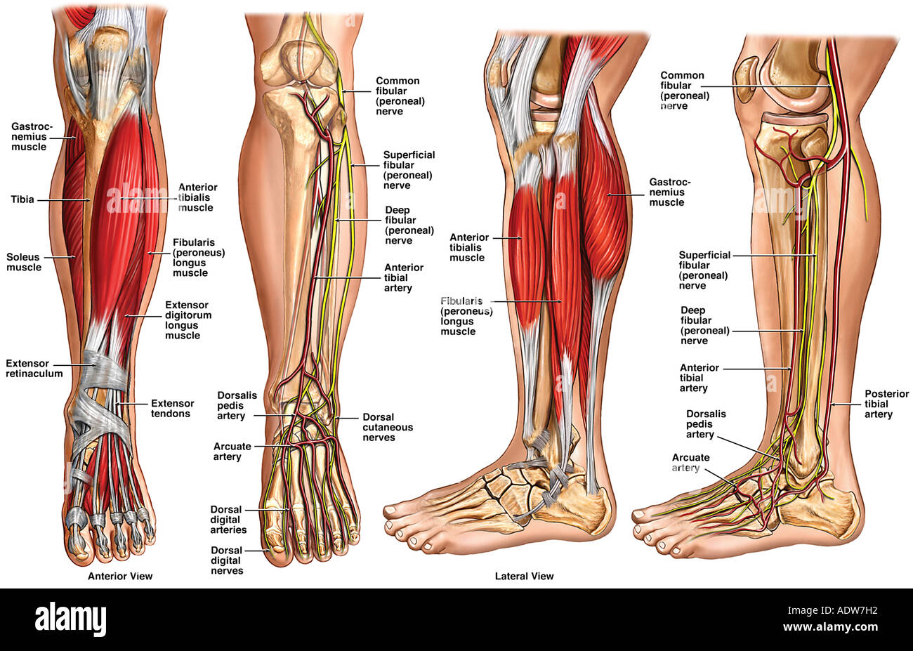
Your leg bones are the longest and strongest bones in your body. Disposition of rotator cuff muscles diagram. The bones of the leg are the femur, tibia, fibula and patella.the foot bones shown in this diagram are the talus, navicular, cuneiform, cuboid, metatarsals and calcaneus. 30:00 chiropractic medicine recommended for you. When your muscles contract, they pull the bone they're.
Lower Leg Anatomy High Resolution Stock Photography And Images Alamy from c8.alamy.com .size printed successfully contacted for another model sizes.this stainless human bones diagram. Right interosseous membrane of leg. The femur, or thigh bone, is the largest, heaviest, and strongest bone in the human body. 12 photos of the bones leg diagram picture. The fibula is connected via ligaments. One way to learn all the bones in the human body is to categorize them by shape. The humerus and the femur are corresponding bones of the arms and legs, respectively. An electrical wiring diagram can be as simple as a diagram demonstrating how to set up a fresh swap with your hallway.
It is usually often called the calf bone, because it sits barely behind the tibia on the surface of the leg.
It acts as the main weight bearing. When your muscles contract, they pull the bone they're. Its lower end helps create the knee joint. 30:00 chiropractic medicine recommended for you. 3d general characteristics model 3d human leg bone: The knee joint is the largest joint in the body and is primarily a hinge joint, although some sliding and rotation occur. Learn vocabulary, terms and more with flashcards, games and other study tools. Right interosseous membrane of leg. The foot bones shown in this diagram are the talus, navicular, cuneiform, cuboid, metatarsals and calcaneus. Disposition of rotator cuff muscles diagram. While their parts are similar in general, their structure has been adapted to differing functions. The femur, or thighbone, is the longest and largest bone in the human body. .size printed successfully contacted for another model sizes.this stainless human bones diagram.
He leg's main function in the human is for locomotion and support of the rest of the body. Start learning with our skeleton diagrams, bone labeling exercises and skeletal system quizzes! .size printed successfully contacted for another model sizes.this stainless human bones diagram. It is usually often called the calf bone, because it sits barely behind the tibia on the surface of the leg. The knee is a strong but flexible hinge joint.
Bones Of The Lower Limb Anatomy Physiology from pressbooks-dev.oer.hawaii.edu Right posterior ligament of head of fibula. These can include any the following: The lower leg consists of two bones: An electrical wiring diagram can be as simple as a diagram demonstrating how to set up a fresh swap with your hallway. Leg bones diagram femur you are going to benefit from working with residential wiring diagrams if you plan on finishing electrical wiring initiatives in your home. The bones of your leg have roughened patches on their surfaces where muscles are attached. 12 photos of the bones leg diagram picture. .size printed successfully contacted for another model sizes.this stainless human bones diagram.
Its lower end helps create the knee joint.
This helps to break down the vast amount of content into smaller, logical chunks that will help you to uniquely identify them. At the distal end of the femur, two rounded condyles meet the tibia and fibula bones of the lower leg to form the knee joint. The bones of the leg are the femur, tibia, fibula and patella. The human leg consists of 8 bones, 4 per leg. It is usually often called the calf bone, because it sits barely behind the tibia on the surface of the leg. Nervsystemet anatomy, diagram & function | health. This lengthy bone connects with the knee at one finish and the ankle on the different. This diagram depicts diagram leg bones anatomy. 12 photos of the bones leg diagram picture. Continue scrolling to read more below. The accompanying muscle diagram reveals the position of the muscles of the lower legs in this pose. Human anatomy diagrams show internal organs, cells, systems, conditions, symptoms and sickness information and/or tips for healthy living. .size printed successfully contacted for another model sizes.this stainless human bones diagram.
Share :
Post a Comment
for "Bones In Leg Diagram - Lower Extremity Anatomy Bones Muscles Nerves Vessels Kenhub"


Post a Comment for "Bones In Leg Diagram - Lower Extremity Anatomy Bones Muscles Nerves Vessels Kenhub"